Radiology
The Fillmore County Hospital Imaging Department provides a wide range of diagnostic and interventional services. The department encompasses both radiological and ultrasound procedures. All exams require orders from a medical provider.
Hours of Operation
Monday through Friday from 7:00 am to 5:00 pm
Available 24/7 for emergency needs
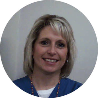
Kim Taylor, RT(R)(M)(CT)
Radiology manager
(402) 759 – 3167 ext 400
ktaylor@myfch.org
CT Scan
MRI
Fluoroscopy
PET/CT
Nuclear Medicine
Ultrasound
X-Ray
X-ray is not used just for detecting abnormalities in the bones. It can be used to image the chest to look for possible diseases/conditions of the heart and lungs. It may also be used to look at the abdomen to image abnormalities that can include kidney stones or blockages in the bowel.
Here at Fillmore County Hospital we offer a variety of services performed by an interventional radiologist. Interventional radiologists are considered the surgeons of radiology as they able to treat patients will minimally invasive procedures with the use imaging guidance. Often these procedures are performed with less pain and faster recovery times. Many common procedures we offer at Fillmore County Hospital include joint injections, injections for back pain, Tenex procedures, treatments for migraines, and numerous other procedures.
- Joint Injections: A joint injection of the shoulder and hip are common sites of arthritis. A combination of anesthetic and steroid are utilized to decrease pain in these joints. The patient is brought to the x-ray room where the site of pain is prepped in a sterile fashion. Live x-rays are used to guide a needle into the joint. An iodinated contrast is then used to confirm the position, and the medicine is injected. The typical time taken for this injection can be 10-30 minutes.
- Injections for Back Pain: There are different variations of back injections used to treat specific types of pain caused by either nerves or arthritis in the back. The interventional radiologist uses a combination of anesthetic and steroid to treat theses sites. A C-arm, live x-rays, is used to find the desired angle in the spine to help the interventional radiologist direct the medications into the affected area. Patients are required to have a driver accompany them as there is a chance of numbing in the legs. A patient can expect to be at the hospital for 45-60 minutes from the check-in process to the check-out process.
- Tenex Procedures: Tenex Health TX™ was designed in collaboration with MAYO Clinic. With the assistance of ultrasound, the interventional radiologist will identify the location of damaged tissue. A small incision is then made to insert the tip of the Tenex machine. Ultrasound guidance is used through out the procedure to ensure only damaged tissue is extract while maintaining healthy tissue. Typical sites the Tenex is used include the foot for plantar fasciitis and the elbow for “tennis” elbow.
- Migraine & Headache Treatment: The SphenoCath procedure is an effective tool in treating migraines, facial pain, cluster headaches and trigeminal neuralgia. Medication is administered via the nasal passages to provide immediate relief. This is a needle-free procedure that takes just a few minutes to perform. We have treated a variety of ages ranging from pediatrics on up.
Mammography
In September of 2019, Fillmore County Hospital upgraded their mammography unit to include 3D. This new technology provides more information for the radiologist to interpret the breast. Some of the benefits include better imaging of dense breast tissue, earlier detection of breast cancers, and a reduced chance of being called back for additional views. Mammography is offered Monday through Friday at Fillmore County Hospital.
Screening mammograms allow radiologists to find breast abnormalities even before they can be felt during a physical exam. The successful treatment of breast cancer is often linked to early detection and diagnosis. FCH uses 3D mammography (also known as Digital Breast Tomosynthesis or DBT), the new standard for annual screening, that is covered by all major insurance companies.
Diagnostic mammograms are used to evaluate a patient with abnormal symptoms such as a breast lump, nipple discharge or “cystic” breasts that may have been noticed during an exam or screening mammogram.
For women who have no health insurance or other circumstances that keep them from having a screening mammogram, there are resources available for Breast Health Services for participants who qualify.
Through the state of Nebraska – Every Woman Matters Program: for more information, go here.
Through Blue Valley Community Action Health Services – Bridge to Care:
Breast Tomosynthesis
Genius 3D Mammography
Osteoporosis
There are three types of bone density: normal, osteopenia, and osteoporosis. As we age, our bone density begins to decrease.
Osteopenia vs Osteoporosis
Osteopenia is a condition of the bone that is weaker/less dense than normal bone. Its classification sits between normal and osteoporosis. On average, osteopenia begins in your 50s. Treating osteopenia early may help slow the progression of bone loss that leads to osteoporosis.
Osteoporosis is when your bones become more porous and lose density. When this occurs, the likelihood of a fracture increases greatly. Two common signs of osteoporosis are a hunched back and height loss. Osteoporosis is a common risk for hip fractures, wrist fractures, and compression fractures of the spine. When the bones become more porous and brittle, even something as simple as a sneeze or minor fall can lead to a fracture.
Prevention
There are numerous ways to help prevent or slow down the progression of osteopenia and/or osteoporosis. Many include:
- Exercise that includes strength or resistance training
- Taking a calcium supplement with Vitamin D
- Eating foods rich in Vitamin D and calcium: spinach, broccoli, leafy greens, non-fat or low-fat yogurts and cheeses, and pink salmon
- Cessation of smoking
Osteoporosis can not be reversed. Bone Density Exams are important in detecting the disease early so you and your provider can make a plan to slow the progression of bone loss. Fillmore County Hospital offers bone density exams, DXA, Monday through Friday. Call your provider to see if you are eligible to have your bone density exam.